Lab Test Results of HIV inactivation by electric current from Appendix E
Appendix 1.
Lab Test Results of HIV inactivation by electric current from Appendix E Paper by W. Lyman, et al. Reporting Inactivation of AIDS Virus by Electric Current
William D. Lyman, Irwin R.Merkatz William C. Hatch and Steven C. Kaali Departments of Pathology, and Obstetrics & Gynecology Albert Einstein. College of Medicine, 1300 Morris Park Ave., Bronx, N.Y.10461
William D. Lyman, Irwin R.Merkatz William C. Hatch and Steven C. Kaali Departments of Pathology, and Obstetrics & Gynecology Albert Einstein. College of Medicine, 1300 Morris Park Ave., Bronx, N.Y.10461
SUMMARY
In this report, we present the results of double-blinded studies on the use of direct electric current to alter the infectivity o£ HIV-1 for susceptible cells in vitro. Two lymphoblastoid cell lines (H9 and CEM-SS) were exposed to aliquots of the RT strain of HIV-1 treated with direct current. Results of these studies show that virus treated with currents from 50 to 100 microamperes (ìA) has a significantly reduced infectivity for susceptible cells.
These experimental currents were equal to 3.85 and 7.7.ìÁ/mmcurrent densities respectively. The reduction of infectivity was dependent upon, the total electric charge (ìA x min) passing through the chamber to which the virus was exposed. Viral infectivity was determined by two independent measures: a syncytium-formation assay which can be used to quantify the production of infectious particles; and. a reverse transcriptase assay which is an index of viral protein production. Additional experiments demonstrated that the currents employed were biocompatible. Uninfected H9 cells were exposed to the same conditions used for the viral aliquots.
There was no significant change in the percentage of viable uninfected cells exposed to any of the currents tested. Therefore, because biocompatible direct electric current attenuates the infectivity of cell-free virus, this treatment may allow development of new strategies to prevent transmission of HIV-1 through either treating the general blood supply or developing alternative barrier contraceptive devices. Additionally, biocompatible electric. current may be applicable for the direct treatment of AIDS patients by utilizing either extracorporeal systems or self contained indwelling electrodes. 15
Lastly, because the virus is being attenuated, electric current may also render treated HIV-1 suitable for vaccine development.
Key words: HIV-1, AIDS, treatment, suppression of infectivity, electricity
INTRODUCTION
The number of individuals infected by the human immunodeficiency virus type-1 (I-(HIV-1) continues to increase on a world-wide basis (1). A significant percentage, if not all, of these individuals will eventually develop the acquire d immunodeficiency syndrome (AIDS) (2)- While horizontal transmission in the homosexual. population may be contained or decreasing (3), heterosexual transmission and infection through contaminated blood supplies continues to increase (4). Additionally ver tical transmission from infected females to their fetuses is also on the rise with a resultant increase in the number of children with AIDS (5). New strategies, therefore, must be devised in order to limit more effectively the spread of this virus.
In this regard, three principal approaches are currently being investigated. In order to decrease susceptibility to the consequences of infection, vaccines are being sought which will induce the production of protective antibodies (6). As treatment modalities, the use of soluble antagonists to block the receptor for HIV-1 is being studied (7) as are pharmacologic agents such as nucleic acid analogs which can interfere with the transcription of viral genomic sequences (8). Each of these systems has------------ and limitations and to date none has proven completely effective.
Because heat or light in combination with drugs and dyes can inactivate viruses including HIV-2 in vitro (9), others have suggested the use of these forms of energy to treat .. AIDS patients. The results of studies using heat have not been peer- reviewed and are therefore impossible to evaluate. The use of light with drugs ["photopheresis"] (10) appears to be efficacious although this treatment may be limited by drug toxicity and the potential long-term effects of ultraviolet radiation on blood c ell nucleic acids. Also, by its nature, this last system may not be suitable for the treatment of tissue-associated virus.
As result of our interest in the use of electric current to alter biological systems , we focused our investigations on the ability of direct electrical current at biocompatible levels to alter the infectivity of HTV-1 for susceptible CD4 positive cell s in vitro.
MATERIALS AND METHODS
Electrical treatment of HIV 1:
The RF strain of HIV-I (AIDS Reagent Program) was cryopreserved prior to treatment at -70°C. Fur treatment, a sample of virus was thawed and maintained on ice at 4°C . Ten microliters (ìl) of HIV-1 at a concentration of 105 infectious particles per ml were placed into a chamber which included a pair of platinum electrodes 1mm apart permanently mounted into a well 1.56mm in length an d 8.32mm in depth equal to 12.9 ìl volume capacity. The chamber was connected to a power supply capable of creating constant direct current. The viral aliquots were exposed to direct currents ranging from 0 microamperes ( ìA) for up to 12 minutes to 100ìA for up to 6 minutes. Intermediate currents of 25, 50 and 75ìA were used to expose similar viral aliquots. Under these conditions, for example, 0, 50 and 100ìA represent 0, 3.85 and 7 .7ìA/mmcurrent densities respectively. The current was monitored throughout the experiment. A matrix of current and time employed is shown in Table 1.
After the exposure of virus to electric current, the contents of the chamber were removed and placed into sterile microtubes. Five ìl of each sample were removed and diluted with 95ìl tissue culture medium supplemented with 10% fetal calf serum (FCS) for subsequent assays.
Syncytium-formation assays:
This assay was performed as previously described by Nara et al (11). Briefly, 10CEM-SS cells were dispensed into poly-L-lysine coated microliter wells. Thereafter, tenfold dilutions o f H9 cells incubated with the treated HIV-1 samples were co-cultured in triplicate for up to 4 days with the CEM-SS cells. Identical wells were prepared with control uninfected and infected cells. The wells were examined for syncytium formation at 2 and 3 days and quantified using an inverted microscope.
Reverse trascriptase assay:
Uninfected H9 cells, were pelleted at 1,000 rpm for minutes at room temperature, the supernatant was decanted and the cells were resuspended in 100ìl treated viral sample. The cells were incubated for up to 6 hours with the viral samples. At the end of the incubation time, the viral/cell suspensions were centrifuged at 1,000 RPM for 5 minutes and the supernatant decanted. The cell pellet was then resuspended in 5ml of RPMI, 10% FCS and placed into a T25 tissue culture flask and maintained at 37C, 5% COin a humidified chamber. At 2 day intervals (beginning at day 21ml of the cell suspensions was removed from each sample and centrifuged at 1,000 rpm for 5 minutes in order to pellet the cells. The supernatant was subsequently centrifuged at 14,000 RPM for I5 minutes. The pellet was resuspended in suspension buffer and assayed using standard methodology employing Mg+ + as the divalent cation poly (rA) oligo d(T) 12-18 as template primer, and tritiated thymidine (H-TdR) which comprise the reaction mixture. 17
Known HIV positive and negative control samples were included in each assay for reference. Thirty ìl of the reacti on mixture were added to each 10 ìl viral sample and incubated at 37 C for 60 min. Samples were then incubated with 1ml of cold quench solution on ice for 15 minutes and filtered through a Millipore manifold. Chimneys were rinsed first with wash solution and followed by cold 95% ethanol. The filters were dried by vacuum and counted in scintillation fluid. Reverse transcriptase activity is expressed as counts per minute (cpm) and is considered positive only if cpm are at least five times greater than the cpm obtained with HIV negative control samples.
Biocompatibility of electric currents/time:
To determine if the electric currents used were in a biocompatibility range of energy, uninfected H9 cells were exposed to distinct currents for different amounts of time. The H9 cells were washed two times in Hanks Balance Salt Solution (HBSS). Thereafter, the cells were resuspended in RPMI, 10% FCS at a concentration of 10cells per ml, Ten ìl of the cell samples were placed into the reaction c hamber. The cell samples were then exposed to 0, 50 or 100ìA for 0, 3 or 6 minutes. At the end of each test, the cell sample was removed from the chamber and approximately 10ìl of the sample was mixed with 90ìl of trypan blue. The number of viable cells w as determined by trypan blue exclusion using a hemocytometer and tight microscope. Results are expressed as percentage of viable cells from the total of all cells. At least 200 cells per field were counted.
Statistical analysis:
Results of the syncytium-formation and reverse transcriptase assays were tested for statistical significance by the Student's t test and analyses of variance.
RESULTS
Syncytium-formation assay:
Using this index of HIV-1 infectivity, it was determined that exposing virus to direct electric current suppressed its capacity to induce the formation of syncytia. Figure 1 shows a representative e xperiment and Table 2 shows the Croup data for 3 separate experiments. As can be noted in Figure l, a statistically significant (p<0.001) reduction in sycytium number was observed and this reduction was dependent upon the current applied to the viral i solate. At three different viral dilutions, there were analogous results in that a total charge of 200ìA x min (25ìA for 8 minutes) reduced the number of syncytia from 50 to 65% while a charge of 300ìA x min (50ìA for 6 minute s, 75ìA for 4 minutes or 100ìA for 3 minutes) resulted in 90% reduction. 18
Reverse transcriptase assays:
The direct electric currents to which HIV-1 was exposed also reduced reverse transcriptase activity. Five separate experiments were conducted and a representative experiment is shown in Figure 2 and the ;coup data are included in Table 3. As can be seen in Figure 2, there was a significant decrease in the amount of reverse transcriptase activity after exposure of the virus to either 50ìA for 3 or 6 minutes. An equivalent reduction in reverse transcriptase activity was also noted with exposure to, 100ìA for 3 minutes and almost ablation of reverse transcriptase activity was seen with exposure of the viral isolate to 100ìA for 6 minutes. The group data (Table 3} show that after exposure to 50ìA for 6 minutes, there was a 44% reduction in activity and treatment of virus with 100ìA for 6 minutes resulted in a 94% reduction. An analysis of variance indicates that t he decrease in reverse transcriptase activity was statistically significant (p <0.0001).
Biocompatibility of the electric currents/time:
The results of a viability analysis using trypan blue exclusion criteria applied to uninfected cells exposed to the different currents and times used far these studies are shown in Table 4. The viability of H9 cells, after exposure to 100ìA fur either 3 or b minutes, did not show a significant decrease when compared to the 0 Current control. After maximum treatment at 100ìA for 6 minutes, cell viability was 93%. Interestingly, in other preliminary experiments in which HIV-infected H9 cells were used, the results show that at 100 ìA there may have been a significant decrease in the number of viable cells. That is, while an insta ntaneous pulse of 100 ìA did not affect the viability of infected cells, at 3 and 6 minutes of exposure to 100 ìA, a decrease in viability was noted. This decrease was time dependent in that exposure to 100 ìA far 3 min utes resulted in a viability of 83% while 100 ìA for 6 minutes resulted in a viability of 80%. Although these data are provocative, they only represent a preliminary experiment and require further investigation.
With respect to the possibility that the electric current was transduced into heat, the calculated rise in temperature within the chamber was determined to be less than 1°C. In order to verify this, a temperature microprobe was introduced into the cham ber containing tissue culture medium alone. Results of these studies are shown in Table S. Similar results were obtained when H9 cell-containing medium was placed in the reaction chamber. The data indicate that for the currents and times used for these ex periments, there was no alteration in the temperature of the chamber.
DISCUSSION
The results reported here demonstrate that HIV-1 treated with direct electric currents from 50 to 100ìA has a significantly reduced infectivity for susceptible cells in vitro. This reduction o f infectivity correlates with the total electric change passing through the chamber. Although extrapolation of these data predicts that ablation of HIV infectivity may be possible, and additional preliminary data support this prediction, the expectation t hat some virions may still escape the electrical effect cannot be discounted. Nevertheless, the .therapeutic potential of electric current may reside in its ability to lower the viral titer to subclinical significance or in its incorporation into a strate gy analogous to that of other therapies in which repeated cycles of treatment eventually achieve remission or cure.
The data presented in this report are based on both quantitative and quantal determinations of viral infectivity. Although the syncytium-formation assay can be used to quantify the number of infectious viral particles, this use with respect to HI V-1 may be abridged because of the ability of free fusigenic peptide (gp41) to induce syncytia by itself. Therefore, while syncytia were observed at some dilutions of electrically-treated virus, this may simply represent the presence of soluble gp41 in th e tissue culture medium. We believe that the correlation between total charge and reduction in syncytium number more adequately reflects the ability of direct electric current to reduce HIV-1 infectivity.
This belief is also supported by the results of the reverse transcriptase assays.
Although a decrease in HIV-1 reverse transcriptase does not assure reduced infectiousness of this virus for Susceptible cells; we feel that, taken together with the syncytium-formation data, the results indicate that significant attenua tion of HIV-I infectivity is achieved by treatment with direct electric currents.
With respect to the biocompatibility of the electric currents and total charges reported here, two separate sets of evidence are applicable. The first has to do with the results showing that, by trypan blue exclusion, no significant cyt otoxicity was induced in by any total charge tested. The other evidence is obtained from reports which clearly indicates that the amount of electricity used for these experiments is significantly below presently used therapeutic electric currents which ar e in the milliampere range (12-16).
Rather than negative effects, exposure of cells to electric current may actually have positive consequences for resistance to infection in that important cellular electrochemical changes correlate with enhancement of specific enzymatic activities. In particular, a facilitation of succinate dehydrogenase (SDH) and ATPase activity has been observed (12,15). Both of these enzymes are associated with the oxidative capacity of the cell. Specifically, it has been suggested that an elec trochemical reaction occurs between mitochondrial membrane-bound H+ ATPase and ADP leading to the formation of ATP. Therefore, exposure of cells to direct electric current may directly or indirectly increase 20
energy resources within a cell and facil itate cell metabolism. This, in turn, may actualIy render a cell less susceptible to the effects of viral infection.
In summary, the data presented here indicate that biocompatible direct electric current significantly reduces the infectivity of HIV-1. Continuing investigations are exploring the mechanisms through which this effect is mediated. The in itial focus of these experiments is centered on the potential role which ionic and molecular species generated by electrolysis may have on the virus. However, the complete mechanism by which direct electric current attenuates HIV-1 infectivity is undoubte dly far re complex than simple electrolysis. Nonetheless. and independent of a complete understanding of all of the mechanisms involved in the attenuation of HIV-1 infectivity, the present observations may serve as an initiaI step for the development of new strategies to treat infection or prevent transmission of HIV-1 through either treat ing the general blood supply or developing alternative barrier contraceptive devices. It may also be feasible to treat AIDS patients with direct electric current using either extracorporeal systems or self contained indwelling electrodes. Lastly, because viral infectivity is being attenuated, electric current may render treated HIV-1 suitable for vaccine development.
ACKNOWLEDGMENTS
Thanks go to Mrs. Agnes Geoghan for her excellent secretarial assistance and to Dr.Gabor, Kemeny for important technical help. Additional thanks go to Drs. Frank Lilly and Philip Aisen for their constructive criticism of this manuscript.
LEGENDS
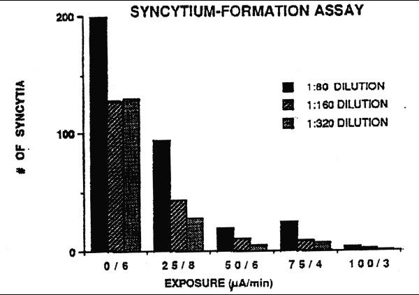
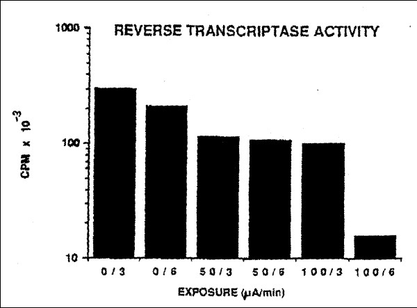
Table 1
Experimental Paradigm
Current (ì.A). Time (Minutes)
0 |
14812 |
25 |
24812 |
50 |
34612 |
75 |
24812 |
100 |
13412 |
Table 2
Effect of ELECTRIC Current on Syncytium Formation a
% of O Current Control (Ä%) b
Current (ìA) Six Minute Exposure
0 |
100 (0) |
|
50 |
50 (-50) |
|
100 |
35 (-65) |
|
a = Value at I:160 dilution of virus. |
||
b = Value equals the mean of 3 experiments. |
||
Table 3
Effect of Electric Current on Reverse Transcriptase Activity a
% of O Current Control (Ä%) Current (ìa) Six Minute Exposure
0 |
100 (0) |
|
50 |
56 (-44) |
|
100 |
6 (-94) |
|
a = Value equals the mean of 5 experiments. The standard error of the mean in each case was less than 10% of the mean value. |
||
Table 4
Effect of Eclectic Current onViability
of Uninfected H9 Cells
(% Viable CeIIs) a
Length of exposure (Minutes),
Current (ìA) 0 3 6
0 |
96946 |
|
50 |
989598 |
|
100 |
969793 |
|
a = At least 200 cells counted in hemocytometer field |
||
Table 5
Effect of Electric Current on Temperature of
Tissue Culture Medium (°C) Length of Exposure (Minutes)
Current (ìA) |
036 |
|
0 |
191919 |
|
50 |
191919 |
|
100 |
191919 |
|
a = The temperature was monitored before, during and after exposure. Results shown are end-point determinations. |
||
REFERENCES
1. Sato PA, Chin J, Mann JM. Review of AIDS and HIV infection Giobal epidermiology and statistics. AIDS 1989; 3 Suppl.1:S301-7.
2. Centers for Disease Control. Revision of the CDC surveillance case definition for acquired immunodeficiency syndrome. MMWR 1987; 1 Suppl. 36:S1-15.
3. Thacker SB, Berkelman RL. Public health surveillance in the United States. Epidemiol. Rev 1988; 10:164.90.
5. Oxtoby MJ. Epidemiology of pediatric AIDS in the United States. In: Brain in Pediatric AIDS (Kozlowski PB, Snider DA, Vietze PM, Wisniewski HM, eds) 1990:1-8
6. Broder S, Mitsuya H, Yarchoan R, Pavlakis GN. Antiretroviral therapy in AIDS. Ann Int Med 1990: 113:604-18.
7. Perno CF, Baseler MW, Broder S, Yarchoan R. Infection of monocytes by human immunodeficiency virus I blocked by inhibitors of CD4-gp120 binding, even in the presence of enhancing antibodies. J Exp Med 1990; I71:1043-56.
8. Mitsuya H, Weinhold KJ, Furman FA et al. 3'-Azido-3'-deoxythymidine (BW A509U): an antiviral agent that inhibits the infectivity and cytopathic effect of human T-lymphotropic virus type III/ lymphadenopathy-associated virus in vitro Proc Natl Acad Sci USA 1985; 82:7096-100.
9. Quinnan GV, Wells MA, Wittek AE, et al. Inactivation of human T-cell virus, type III by heat, chemicals and irradiation. Transfusion 1986; 26:481-3.
10. Bisaccia E, Berger C, KIainer AS. Extracorporeal photopheresis in the treatment of AIDS-related complex: A pilot study. Ann Int Med 1990; 113:270-75.
11. Nara PL, Hatch WC, Dunlop NM, et al.: Simple, rapid quantitative, syncytium-forming microassay for the detection of human immunodeficiency virus neutralizing antibody. Aids Res Hum Retrovirus 1987; 3:283-302
12. Cheng N, Van Hoof H, Bockx E, et al. The effects of electric currents on ATP generation, protein synthesis, and membrane transport in rat skin. Clin Ortho ReI Res 1982; 17I:26472.
13. Frank G, Schachar N, Dittrich D, et al. Electromagnetic stimulation of ligament healing in rabbits. Clin Ortho ReI Res 1983; I75:263-72.
14. Eriksson E, Haggmark T. Comparison of isometric muscle training and electrical stimulation supplementing isometric muscle training in the recovery after major knee ligament surgery. Amer J Sports Med 19?9; 7:159-71.
15. Stanish WD, Valiant GA, Bonen A, et al. The effects of immobilization and of electrical stimulation on muscle glycogen and myofibrillar ATPase. Can J Appl Sport Sci 1982; 7:267-71.'
16. Pills AA. Electrochemical information transfer at living cell membranes. Ann NY Acad Sci 1974; 205:148-70.