Observations with the Rife Microscope
Microscope
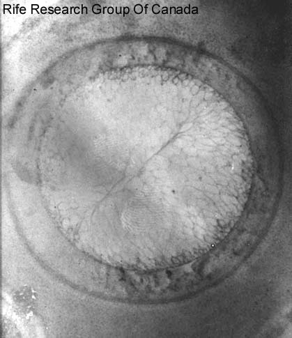
Dr. Edward C. Rosenow, MD (1875-1966) was one of the USA's prominent medical authorities, and was the head of the Mayo Foundation's Experimental Bacteriology program from 1915 to 1944. His son, Edward Jr., was also a prominent physician, and from 1959 to 1977 was Director of the American College of Physicians. It is of interest that Edward Jr. was practicing Internal Medicine in Pasadena, CA. in the late 1930's. After the report, you can see a selection of images made with the original Rife microscope, supplied by the Rife Research group in Canada.
Observations with the Rife Microscope of Filter-Passing Forms of Microorganisms
by Dr. Edward C. Rosenow, Rochester, Minnesota, USA
Recently, I reported to the staff of the Mayo Clinic the more important observation made during three days, July 5, 6 and 7, 1932, spent in Dr. Kendall's laboratory at Northwestern University Medical School, Chicago. I went there at the invitation of Drs. Kendall and Rife, to share with them their observations in a restudy of the filter-passing forms of Eberthella typhi as seen with an improved model of the Rife microscope. They asked me also to bring with me my cultures of the streptococcus from poliomyelitis.
I would like to repeat here that portion of my report which had to do specifically with the Rife microscope.
Owing to the novel and important character of the work, each of us verified at every step the results obtained. Microscopic examinations of suitable specimens was made as a routine by Dr. Rife with his high-power microscope, by Dr. Kendall with the oil immersion dark field, and by myself with the ordinary Zeiss microscope equipped with a 2 mm apochromatic oil immersion lens and x10 ocular giving a magnification of about 900 diameters. Most observations with the Rife microscope were made at 8,000 diameters. In order to check the magnification, gram and safranin stained films of cultures of Eberthella typhi, of the streptococcus from poliomyelitis, and stained films of blood, and of the sediment of the spinal fluid from a case of acute poliomyelitis were examined. Bacilli, streptococci, erythrocytes, polymorphonuclear leukoeytes and lymphocytes were clearly seen, and in each instance were, as nearly as could be estimated, about nine times the diameter as when examined with the 2 mm oil immersion at about 900 diameters.
The following principles and methods were stated by Dr. Rife as being essential in order to visualize clearly the objects at this and higher magnifications by direct observation. Spherical aberration is reduced to the minimum and magnification greatly increased by using objectives in place of oculars. Proper visualization, especially of unstained objects, is obtained by the use of an intense beam of monochromatic polarized light created by rotating wedge-shaped quartz prisms placed between the source of light and the substage quartz condenser. Dispersion of the transmitted rays of light, as they pass upward to the eye, is prevented by passing them through a series of quartz erecting (90 degrees) prisms. Projection of the rays of light through air is not greater than 30 mm at any point.
In my original report (Proc. Staff Meeting Mayo Clinic, 7: 408-413 July 13, 1932) I summarized as follows:
"There can be no question of the existence of the filterable turquoise blue bodies of Eberthella-typhi described by Kendall. They are not visible by ordinary methods of illumination and magnification, not because they are too small, but rather, it appears, because of their peculiar non-staining hyalin structure. Their visualization under the Rife microscope is due to the ingenious methods employed rather than to excessively high magnification. Examination under the Rife microscope of specimens, containing objects visible with the ordinary microscope, leaves no doubt of the accurate visualization of objects or particulate matter by direct observation at the extremely high magnification (calculated to be 8,000 diameters) obtained with this instrument."
The findings under the Rife microscope of cocci and diplococci in filtrates of cultures of the streptococcus from poliomyelitis, and in filtrates of the viruses of poliomyelitis and herpes encephalitis, not detectable by the ordinary methods of examination, and which resembled in form and size those found in the respective cultures, and the absence of minute forms, suggest that the filterable, inciting agent of these diseases is not necessarily extremely small, as is universally believed. Indeed, the filterable, inciting agent may be the non-staining, highly plastic, hyaline stage of the visible, stainable, cultivable organism, the streptococcus.
It is, of course, possible that these unstained, invisible forms revealed by ordinary methods of examination are not the inciting agents or 'viruses' of these diseases and that they represent merely the filterable or other state of the streptococcus. A consideration of the great difficulty one has in isolating the streptococcus and demonstrating diplococci in lesions in these diseases and the ease with which the bodies are found in the filtrate indicate clearly that the `invisible' forms of the streptococcus, if such they be, are present in large numbers in the host, as in positive cultures of the streptococcus. Their form, size and color are too characteristic and true to type to permit considering them as artifacts or as being expressive of etiologically unrelated, contaminating streptococci. Non-infectivity of the filter-passing forms, except in the cases of virus diseases, their presence in large numbers in filtrates, both of cultures and of infected tissues, and the great difficulty in obtaining the visible forms in cultures of filtrates indicate that "invisible," filter-passing forms represent a certain stage in the development of microorganisms.
Dr. Edward C. Rosenow, MD (1875-1966) war eine der führenden medizinischen Autoritäten der USA und leitete von 1915 bis 1944 das Programm für experimentelle Bakteriologie der Mayo Foundation. Sein Sohn, Edward Jr., war ebenfalls ein bekannter Arzt und von 1959 bis 1977 Direktor des American College of Physicians. Interessant ist, dass Edward Jr. in den späten 1930er Jahren als Arzt für Innere Medizin in Pasadena, Kalifornien, tätig war. Nach dem Bericht sehen Sie eine Auswahl von Bildern, die mit dem Original-Rife-Mikroskop gemacht wurden, das von der Rife Research Group in Kanada zur Verfügung gestellt wurde.
Beobachtungen mit dem Rife-Mikroskop von filterdurchlässigen Formen von Mikroorganismen
von Dr. Edward C. Rosenow, Rochester, Minnesota, USA
Kürzlich berichtete ich dem Personal der Mayo-Klinik über die wichtigsten Beobachtungen, die ich während dreier Tage, am 5., 6. und 7. Juli 1932, im Labor von Dr. Kendall an der Northwestern University Medical School in Chicago gemacht hatte. Ich war auf Einladung von Dr. Kendall und Dr. Rife dorthin gereist, um mit ihnen ihre Beobachtungen bei einer erneuten Untersuchung der filtrierenden Formen von Eberthella typhi zu teilen, die mit einem verbesserten Modell des Rife-Mikroskops beobachtet wurden. Sie baten mich auch, meine Kulturen von Streptokokken aus der Kinderlähmung mitzubringen.
Ich möchte hier den Teil meines Berichts wiederholen, der sich speziell mit dem Rife-Mikroskop befasst.
Aufgrund des neuartigen und wichtigen Charakters der Arbeit hat jeder von uns die erzielten Ergebnisse bei jedem Schritt überprüft. Die mikroskopischen Untersuchungen geeigneter Proben wurden routinemäßig von Dr. Rife mit seinem Hochleistungsmikroskop, von Dr. Kendall mit dem Ölimmersions-Dunkelfeld und von mir selbst mit dem gewöhnlichen Zeiss-Mikroskop durchgeführt, das mit einer apochromatischen Ölimmersionslinse von 2 mm und einem Okular von x10 ausgestattet war und eine Vergrößerung von etwa 900 Durchmessern ergab. Die meisten Beobachtungen mit dem Rife-Mikroskop wurden bei 8.000 Durchmessern durchgeführt. Zur Überprüfung der Vergrößerung wurden gram- und safraningefärbte Filme von Kulturen von Eberthella typhi, von Streptokokken aus Poliomyelitis sowie gefärbte Filme von Blut und vom Sediment der Rückenmarksflüssigkeit aus einem Fall von akuter Poliomyelitis untersucht. Bazillen, Streptokokken, Erythrozyten, polymorphkernige Leukozyten und Lymphozyten waren deutlich zu erkennen, und in jedem Fall waren sie, soweit abschätzbar, etwa neunmal so groß wie bei der Untersuchung mit der 2-mm-Ölimmersion bei etwa 900 Durchmessern.
Die folgenden Prinzipien und Methoden wurden von Dr. Rife als wesentlich bezeichnet, um die Objekte bei dieser und höheren Vergrößerungen durch direkte Beobachtung deutlich sichtbar zu machen. Durch die Verwendung von Objektiven anstelle von Okularen wird die sphärische Aberration auf ein Minimum reduziert und die Vergrößerung stark erhöht. Eine gute Sichtbarkeit, insbesondere von ungefärbten Objekten, wird durch die Verwendung eines intensiven Strahls von monochromatischem, polarisiertem Licht erreicht, das durch die Drehung von keilförmigen Quarzprismen erzeugt wird, die zwischen der Lichtquelle und dem Unterstufen-Quarzkondensor angeordnet sind. Die Streuung der übertragenen Lichtstrahlen auf ihrem Weg nach oben zum Auge wird dadurch verhindert, dass sie durch eine Reihe von Quarzaufrichtungsprismen (90 Grad) geleitet werden. Die Projektion der Lichtstrahlen durch die Luft ist an keinem Punkt größer als 30 mm.
In meinem ursprünglichen Bericht (Proc. Staff Meeting Mayo Clinic, 7: 408-413 July 13, 1932) habe ich das Ergebnis wie folgt zusammengefasst:
"Die Existenz der von Kendall beschriebenen filtrierbaren türkisblauen Körper von Eberthella-typhi kann nicht in Frage gestellt werden. Sie sind mit den üblichen Beleuchtungs- und Vergrößerungsmethoden nicht sichtbar, und zwar nicht, weil sie zu klein sind, sondern vielmehr, wie es scheint, wegen ihrer besonderen, nicht färbenden Hyalinstruktur. Ihre Sichtbarmachung unter dem Rife-Mikroskop ist eher auf die ausgeklügelten Methoden zurückzuführen als auf eine zu starke Vergrößerung. Die Untersuchung von Proben unter dem Rife-Mikroskop, die mit dem gewöhnlichen Mikroskop sichtbare Objekte enthalten, lässt keinen Zweifel an der genauen Sichtbarmachung von Objekten oder Partikeln durch direkte Beobachtung bei der extrem hohen Vergrößerung (berechnet auf 8.000 Durchmesser), die mit diesem Instrument erreicht wird."
Die Entdeckungen unter dem Rife-Mikroskop von Kokken und Diplokokken in Filtraten von Kulturen der Streptokokken von Poliomyelitis und in Filtraten der Viren von Poliomyelitis und Herpes-Enzephalitis, die mit den üblichen Untersuchungsmethoden nicht nachweisbar waren und die in Form und Größe denjenigen ähnelten, die in den jeweiligen Kulturen gefunden wurden, sowie das Fehlen von winzigen Formen lassen vermuten, dass das filtrierbare, auslösende Agens dieser Krankheiten nicht unbedingt extrem klein ist, wie allgemein angenommen wird. Der abfiltrierbare Erreger könnte in der Tat das nicht färbende, hochplastische, hyaline Stadium des sichtbaren, färbbaren, kultivierbaren Organismus, des Streptokokkus, sein.
Es ist natürlich möglich, dass diese ungefärbten, unsichtbaren Formen, die durch gewöhnliche Untersuchungsmethoden aufgedeckt werden, nicht die Erreger oder "Viren" dieser Krankheiten sind, sondern lediglich das filtrierbare oder andere Stadium der Streptokokken darstellen. Eine Betrachtung der großen Schwierigkeiten, die man bei der Isolierung der Streptokokken und dem Nachweis von Diplokokken in Läsionen bei diesen Krankheiten hat, und der Leichtigkeit, mit der die Körper im Filtrat gefunden werden, zeigt deutlich, dass die "unsichtbaren" Formen der Streptokokken, wenn es sich um solche handelt, in großer Zahl im Wirt vorhanden sind, wie in positiven Kulturen der Streptokokken. Ihre Form, Größe und Farbe sind zu charakteristisch und typgerecht, um sie als Artefakte oder als Ausdruck von ätiologisch nicht verwandten, kontaminierenden Streptokokken zu betrachten. Die Nichtinfektiosität der filtrierenden Formen, außer bei Viruserkrankungen, ihr zahlreiches Vorhandensein in Filtraten, sowohl von Kulturen als auch von infiziertem Gewebe, und die große Schwierigkeit, die sichtbaren Formen in Kulturen von Filtraten zu erhalten, deuten darauf hin, dass die "unsichtbaren" filtrierenden Formen ein bestimmtes Stadium in der Entwicklung der Mikroorganismen darstellen.
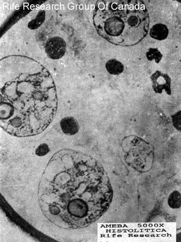
Ameba 5000x Histolitica
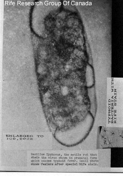
Typhoid Bacteria with Rife Stain
Enlarged to 300,000x
Enlarged to 300,000x
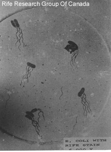
E. Coli with Rife Stain 6000x

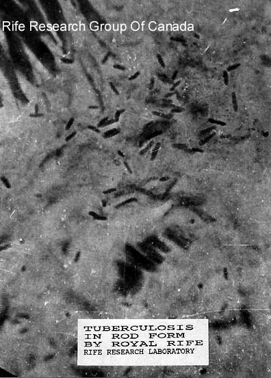
Tuberculosis in rod Form

Strombilitis Intestinalis
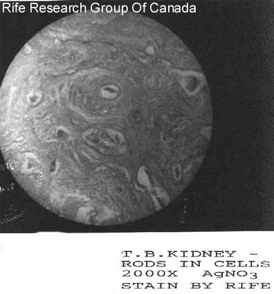
T.B. Kidney - Rods in Cells, 2000x AgNO3 Stain
Tetanus