
Electricity, Frequencies and Cancer
Electricity, Frequencies and Cancer
There have been a number of research papers showing a link between the use of electricity and/or frequencies for the treatment of cancer going all the way back to an article in The Lancet from 1880. We have assembled here a selection of relevant papers for your reference.
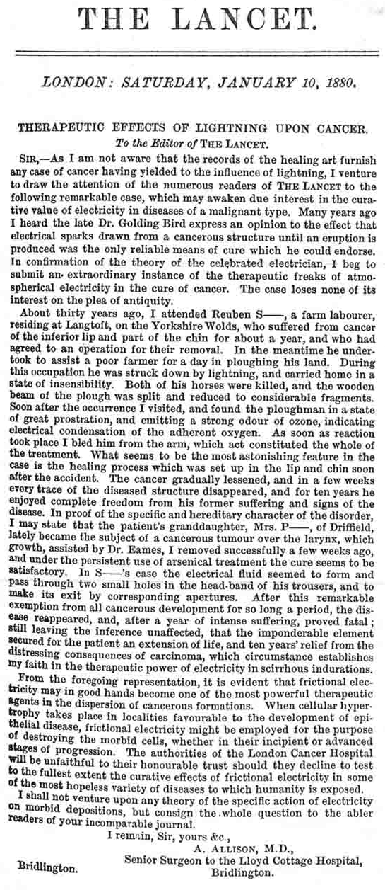
Therapeutic Effects of Lightning Upon Cancer
A. ALLISON, M.D., Senior Surgeon to the Lloyd Cottage Hospital, Bridlington. The Lancet, London: January 10, 1880.
Describes a case where lightning caused a malignant cancer to be cured that was reported in the Lancet.
Ultrasound Cancer Treatment Kills Tumors in Mice
LONDON Jan 29, 2003 (Reuters)
Scientists at a Northern Ireland biotech company used an electric field and ultrasound to kill cancerous cells in mice. Instead of surgery, drugs or radiation treatment, researchers at Gendel used an electric field and to kill cancerous cells in the laboratory and tumors in 50 mice.
 Evaluating Cancer Therapies and Developing a Cancer Program
Don Benjamin, Convenor/Research Officer, Cancer Information & Support Society, St Leonards (Sydney), Saturday 3 May 2003
The medical profession has a good track record in trauma intervention. It does not have such a good record in the treatment of degenerative diseases such as cancer, coronary heart disease and arthritis.
In this presentation I will cover:
The evidence for efficacy of orthodox and alternative therapies
What this means in terms of paradigms for cancer
What is the alternative paradigm?
What therapies fit into this approach?
What is the role of orthodox therapies?
How to put together a cancer control program
Evaluation of DC Current Therapy in Mammary Cancer Tumor
MARCOS TELLÓ, LUCIANA OLIVEIRA DE OLIVEIRA, ROSEMARI TERESINHA DE OLIVEIRA, GUILHERME DIAS, ADROALDO RAIZER
The aim of this paper is to present the results attained by our research group from the clinical applications of DC current in dogs presenting mammary tumors. Indeed, it is important to point out that the cytopathological exams, attained from the treated mammary glands, indicate that the macrophage cells appear in all treated tumors after some application sessions.
Keywords – ElectroChemical Therapy, DC current, mammary glands, cancer.
Pulsed Field Assisted Chemotherapy
James E. Bare, 8005 Marble Ave. NE, Albuquerque, NM 87110
Research into the physiologic effects of low power, pulsed electromagnetic (EM) fields, has produced a number of important discoveries. To date, with rare exception, these discoveries at best are investigational, and have not been applied in a clinical manner. Much of this research material is unknown to the general practitioner, and has not been correlated into a potentially utilizable treatment method. This paper proposes the fusion of existing cancer chemotherapy techniques with low power pulsed EM field research discoveries. Evidence is presented that the sum of these combined effects far exceeds that of each method individually. A transmitted, pulsed EM field, can be created which will safely produce whole body permeation/saturation. Such saturation can create an interaction of the pulsed EM field with chemotherapeutic medications, simultaneously, at all tumor sites throughout the body. By creating a synergism of biochemical, electrochemical , and electronic principles, the practitioner should be able to achieve a superior treatment outcome.
Disruption of Cancer Cell Replication by Alternating Electric Fields
Department of Biomedical Engineering, NovoCure Ltd., Haifa, Israel; Rappaport Faculty of Medicine, Technion—Israel Institute of Technology, Haifa, Israel; Department of Molecular Cell Biology, Weizmann Institute of Science, Rehovot, Israel; and Elisha Medical Centre, Haifa, Israel - Published: CANCER RESEARCH 64, 3288–3295, May 1, 2004
Low-intensity, intermediate-frequency (100–300 kHz), alternating electric fields, delivered by means of insulated electrodes, were found to have a profound inhibitory effect on the growth rate of a variety of human and rodent tumor cell lines (Patricia C, U-118, U-87, H-1299, MDA231, PC3, B16F1, F-98, C-6, RG2, and CT-26) and malignant tumors in animals. This effect, shown to be nonthermal, selectively affects dividing cells while quiescent cells are left intact. These fields act in two modes: arrest of cell proliferation and destruction of cells while undergoing division. Both effects are demonstrated when such fields are applied for 24 h to cells undergoing mitosis that is oriented roughly along the field direction. The first mode of action is manifested by interference with the proper formation of the mitotic spindle, whereas the second results in rapid disintegration of the dividing cells. Both effects, which are frequency dependent, are consistent with the computed directional forces exerted by these specific fields on charges and dipoles within the dividing cells. In vivo treatment of tumors in C57BL/6 and BALB/c mice (B16F1 and CT-26 syngeneic tumor models, respectively), resulted in significant slowing of tumor growth and extensive destruction of tumor cells within 3–6 days. These findings demonstrate the potential applicability of the described electric fields as a novel therapeutic modality for malignant tumors.
Tumor blood flow modifying effects of electrochemotherapy: a potential vascular targeted mechanism
Institute of Oncology Ljubljana, University of Ljubljana, Faculty of Electrical Engineering, Ljubljana, Slovenia - Published: Radiol Oncol 2003; 37(1): 43-8.
The aim of this study was to determine the tumor blood flow modifying, and potential vascular targeted effect of electrochemotherapy with bleomycin or cisplatin.
Materials and methods. Electrochemotherapy was performed by application of short intense electric pulses to the tumors after systemic administration of bleomycin or cisplatin. Evaluated were antitumor effectiveness of electrochemotherapy by tumor measurement, tumor blood flow modifying effect by Patent blue staining technique, and sensitivity of endothelial and tumor cells to the drugs and electrochemotherapy by clonogenicity assay.
Results. Electrochemotherapy was effective in treatment of SA-1 tumors in A/J mice resulting in substantial tumor growth delay and also tumor cures. Tumor blood flow reduction following electrochemotherapy correlated well with its antitumor effectiveness. Virtually complete shut down of the tumor blood flow was observed already at 24 h after electrochemotherapy with bleomycin whereas only 50% reduction was observed after electrochemotherapy with cisplatin. Sensitivity of human endothelial HMEC-1 cells to electrochemotherapy suggests a vascular targeted effect for electrochemotherapy in vivo with bleomycin as well as with cisplatin.
Conclusion. These results show that, in addition to direct electroporation of tumor cells, other vascular targeted mechanisms are involved in electrochemotherapy with bleomycin or cisplatin, potentially mediated by tumor blood flow reduction, and enhanced tumor cell death as a result of endothelial damage by electrochemotherapy.
Key words: sarcoma experimental - drug therapy - blood supply; bleomycin; cisplatin; electroporation
Acid-mediated tumor invasion: how does vasculature affect the growth characteristics?
B. S. Govindan, W. B. Spillman, Jr., J. L. Robertson and W. R. Huckle, Virginia Polytechnic Institute and State University
We study the growth of an implanted avascular tumor in rats, in two-dimensions, based on a model where the mechanism of invasion is centered on tumor-induced acidification of the micro-environment and consequent death of normal cells. The spatial distribution of the acid density around the tumor is found using mean-field analysis. By assuming that the viability of both normal and tumor cells falls sharply below certain threshold values of the local pH, we determine the conditions for the formation and radius of a necrotic core at the center, as a function of the tumor radius. We show mathematically that the mean micro-vessel density (MVD) plays a pivotal role in determining the growth characteristics of the tumor. When the MVD is sufficiently small, accumulation of excess acid inside the tumor leads to the formation of a necrotic core, which occupies a significant fraction of the total area in large tumors. However, necrosis is reduced when the mean MVD inside the tumor is larger than outside because of the more efficient removal of excess acid. At sufficiently high MVD, necrosis might be absent in the tumor, or confined to small regions mostly devoid of micro-vessels. Quantitative estimates of MVD for these different phases of growth are obtained, and verified using explicit cellular automaton simulations. Recent experimental studies on the correlation between necrosis and MVD support our main conclusions.
Key words: tumor growth, tumor differentiation, necrotic core, micro-vessel density.
Disruption of Cancer Cell Replication by Alternating Electric Fields
Eilon D. Kirson, Zoya Gurvich, Rosa Schneiderman, Erez Dekel, Aviran Itzhaki, Yoram Wasserman, Rachel Schatzberger, and Yoram Palti
Low-intensity, intermediate-frequency (100–300 kHz), alternating electric fields, delivered by means of insulated electrodes, were found to have a profound inhibitory effect on the growth rate of a variety of human and rodent tumor cell lines (Patricia C, U-118, U-87, H-1299, MDA231, PC3, B16F1, F-98, C-6, RG2, and CT-26) and malignant tumors in animals. This effect, shown to be nonthermal, selectively affects dividing cells while quiescent cells are left intact. These fields act in two modes: arrest of cell proliferation and destruction of cells while undergoing division. Both effects are demonstrated when such fields are applied for 24 h to cells undergoing mitosis that is oriented roughly along the field direction. The first mode of action is manifested by interference with the proper formation of the mitotic spindle, whereas the second results in rapid disintegration of the dividing cells. Both effects, which are frequency dependent, are consistent with the computed directional forces exerted by these specific fields on charges and dipoles within the dividing cells. In vivo treatment of tumors in C57BL/6 and BALB/c mice (B16F1 and CT-26 syngeneic tumor models, respectively), resulted in significant slowing of tumor growth and extensive destruction of tumor cells within 3–6 days. These findings demonstrate the potential applicability of the described electric fields as a novel therapeutic modality for malignant tumors.
Mathematical Modelling of Tumour Acidity: Regulation of Intracellular pH
S.D. Webb, J.A. Sherratt and R.G. Fish - Heriot-Watt University, Edinburgh and Velindre Hospital, Cardif, UK
Measurements of extracellular pH (pHe) in vivo have shown that the microenvironment in tumours is more acidic than in normal tissue. However, both human and animal tumour cells have been shown to have an intracellular pH (pHi) on the alkaline side of neutrality (pH 7.1-7.2). This gives rise to a reversed pH gradient between tumours and normal tissue- which implies that cells within solid tumours are capable of maintaining their level of pHi at physiological levels, despite lower than normal levels of pHe. In this paper the authors describe a mathematical model that provides a possible explanation for the altered pH gradient observed in tumours. The authors examine the infuence of changes in the microenvironment on the activity of several membrane based ion transport systems Using qualitative analysis the authors show that the pHi of tumour cells is less sensitive to external pH than for normal cells, because of their increased reliance on the inefficient glycolytic pathway for energy production. It is shown that under aerobic conditions the lactate -/H+ symporter could be the most active exchanger in the regulation of pHi in tumour cells. However, under more hypoxic conditions lactate extrusion is reduced, and so this exchanger has little eon resting pHi in these regions. The authors also consider an extended model which incorporates the transfer of acids from the cytosol into acidic organelles. The model demonstrates that one of the major factors involved in the maintenance of cytosolic pH to physiological levels, despite an acidic extracellular pH in hypoxic areas of tumour tissue (median, 6.9-7.0), is enhanced sequestration of cytosolic protons into acidic cellular vesicles such as endoplasmic reticulum, golgi, endosomes and lysosomes.
 Nanosecond pulsed electric fields cause melanomas to selfdestruct
Richard Nuccitelli, Uwe Pliquett, Xinhua Chen, Wentia Ford, R. James Swanson, Stephen J. Beebe, Juergen F. Kolb, and Karl H. Schoenbach - Frank Reidy Research Center for Bioelectrics, Old Dominion University - BioElectroMed Corp. - Eastern Virginia Medical School, Norfolk, VA, USA
We have discovered a new, drug-free therapy for treating solid skin tumors. Pulsed electric fields greater than 20 kV/cm with rise times of 30 ns and durations of 300 ns penetrate into the interior of tumor cells and cause tumor cell nuclei to rapidly shrink and tumor blood flow to stop. Melanomas shrink by 90% within two weeks following a cumulative field exposure time of 120 µs. A second treatment at this time can result in complete remission. This new technique provides a highly localized targeting of tumor cells with only minor effects on overlying skin. Each pulse deposits 0.2 J and 100 pulses increase the temperature of the treated region by only 3 °C, ten degrees lower than the minimum temperature for hyperthermia effects.
Keywords
Skin cancer; Cancer therapy; Tumor; Pulsed electric fields; Pyknosis; Inhibiting angiogenesis; DNA; Nucleus
Electric fields have potential as a cancer treatment
Johanna Miller - Physics Today, www.physicstoday.org, August 2007
Healthy cells have regulating mechanisms that generally limit how rapidly they can divide. Skin cells, for example, normally divide about once every 30 days, but they can divide faster in response to a wound that needs healing. Cancer, however, is characterized by cell division that has gone out of control. In cancer cells, the mechanisms that regulate division break down, and cells spend less time in the quiescent state and more time dividing.
Amplitude-modulated electromagnetic fields for the treatment of cancer: Discovery of tumor-specific frequencies and assessment of a novel therapeutic approach
Alexandre Barbault, Frederico P Costa, Brad Bottger, Reginald F Munden, Fin Bomholt, Niels Kuster and Boris Pasch - Journal of Experimental & Clinical Cancer Research 2009, 28:51 doi:10.1186/1756-9966-28-5
Purpose: Because in vitro studies suggest that low levels of electromagnetic fields may modify cancer cell growth, we hypothesized that systemic delivery of a combination of tumor-specific frequencies may have a therapeutic effect. We undertook this study to identify tumor-specific frequencies and test the feasibility of administering such frequencies to patients with advanced cancer.
Patients and methods: We examined patients with various types of cancer using a noninvasive biofeedback method to identify tumor-specific frequencies. We offered compassionate treatment to some patients with advanced cancer and limited therapeutic options.
Results: We examined a total of 163 patients with a diagnosis of cancer and identified a total of 1524 frequencies ranging from 0.1 Hz to 114 kHz. Most frequencies (57–92%) were specific for a single tumor type. Compassionate treatment with tumor-specific frequencies was offered to 28 patients. Three patients experienced grade 1 fatigue during or immediately after treatment. There were no NCI grade 2, 3 or 4 toxicities. Thirteen patients were evaluable for response. One patient with hormone-refractory breast cancer metastatic to the adrenal gland and bones had a complete response lasting 11 months. One patient with hormone-refractory breast cancer metastatic to liver and bones had a partial response lasting 13.5 months. Four patients had stable disease lasting for +34.1 months (thyroid cancer metastatic to lung), 5.1 months (non-small cell lung cancer), 4.1 months (pancreatic cancer metastatic to liver) and 4.0 months (leiomyosarcoma metastatic to liver).
Conclusion: Cancer-related frequencies appear to be tumor-specific and treatment with tumor-specific frequencies is feasible, well tolerated and may have biological efficacy in patients with advanced cancer.
Trial registration: clinicaltrials.gov identifier NCT0080533
Alternating electric fields (TTFields) inhibit metastatic spread of solid tumors to the lungs
Eilon D. Kirson, Moshe Giladi, Zoya Gurvich, Aviran Itzhaki, Daniel Mordechovich, Rosa S. Schneiderman, Yoram Wasserman, Bernhard Ryffel, Dorit Goldsher, Yoram Palti - This article is published with open access at Springerlink.com
Tumor treating fields (TTFields) are low intensity, intermediate frequency, alternating electric fields used to treat cancerous tumors. This novel treatment modality effectively inhibits the growth of solid tumors in vivo and has shown promise in pilot clinical trials in patients with advanced stage solid tumors. TTFields were tested for their potential to inhibit metastatic spread of solid tumors to the lungs in two animal models: (1) Mice injected with malignant melanoma cells (B16F10) into the tail vein, (2) New Zealand White rabbits implanted with VX-2 tumors within the kidney capsule. Mice and rabbits were treated using two-directional TTFields at 100–200 kHz. Animals were either monitored for survival, or sacrificed for pathological and histological analysis of the lungs. The total number of lung surface metastases and the absolute weight of the lungs were both significantly lower in TTFields treated mice then in sham control mice. TTFields treated rabbits survived longer than sham control animals. This extension in survival was found to be due to an inhibition of metastatic spread, seeding or growth in the lungs of TTFields treated rabbits compared to controls. Histologically, extensive peri- and intra-tumoral immune cell infiltration was seen in TTFields treated rabbits only. These results raise the possibility that in addition to their proven inhibitory effect on the growth of solid tumors, TTFields may also have clinical benefit in the prevention of metastatic spread from primary tumors.
Keywords: Tumor treating fields - Metastases - Immune response
Electromagnetic Signals Are Produced by Aqueous Nanostructures Derived from Bacterial DNA Sequences
Luc Montagnier, Jamal Aissa, Stéphane Ferris, Jean-Luc Montagnier, Claude Lavallée - Interdiscip Sci Comput Life Sci (2009) 1: 81-90
A novel property of DNA is described: the capacity of some bacterial DNA sequences to induce electromagnetic waves at high aqueous dilutions. It appears to be a resonance phenomenon triggered by the ambient electromagnetic background of very low frequency waves. The genomic DNA of most pathogenic bacteria contains sequences which are able to generate such signals. This opens the way to the development of highly sensitive detection system for chronic bacterial infections in human and animal diseases.
Keywords: DNA, electromagnetic signals, bacteria
Excerpt from "Electrical Healing and the Violet Ray"
Gary J. Lockhart, Unpublished book written in 2000, edited by Arthur Lee Jabobson (2007)
This gives an insight into the history of the use of electricity for medical purposes and expands on the lancet article. Electricity was used to reduce weight, grow hair and remove hemorrhoids. In certain instances it restored the sight of nearly blind persons, healed desperate cases of rheumatoid arthritis and removed skin cancer.
Effects of Mechanochemically Activated Doxorubicin and 40 MHz Frequency Irradiation on Human A-549 Lung Carcinoma Cells
Institute of Oncology, Academy of Medical Sciences of Ukraine, Kyiv, UkraineKavetsky Institute of Experimental Pathology, Oncology and Radiobiology, National Academy of Sciences of Ukraine, Kyiv, UkraineInc., Miami, Florida, USA
Aim: To study in vitro influence of mechanochemically activated (MA) doxorubicin (DOXO) and electromagnetic irradiation (EMI) on human lung carcinoma A-549 cells. Methods: Solid state DOXO was MA by input energy 20 W/g during 5 min. Tumor cells were exposed to 40 MHz EMI with power density 2 W/cm at temperature 37 °C. Results: Particles of MA DOXO have sizes 10 time smaller than officinal DOXO, high performance liquid chromatography analysis showed that parameters of officinal and MA DOXO were quantitatively equal. Mechanochemical activation initiated in the drug formation of free radicals with g = 2.005, g = 2.003 and g = 1.97. LD values of MA DOXO were 5 times lower than that of officinal drug. Cell survival decreased in the following way after effects EMI -> officinal DOXO -> MA DOXO -> officinal DOXO + EMI -> MA DOXO + EMI. Conclusion: Treatment by MA DOXO and drug with EMI at 37 °C showed better targeting of drug in human lung carcinoma A-549 cells outcomes than officinal DOXO.
Key Words: A-549 human lung carcinoma cells, doxorubicin, mechanochemical activation, electromagnetic irradiation.
Remote Control for Bacteria - Radio waves switch proteins on and off
Remote-controlled bacteria could be just around the corner. Researchers have found a way to switch cell processes on and off with radio waves.
The following articles can only be read after paying for a subscription service
Therapeutic Electromagnetic Field Effects on Angiogenesis During Tumor Growth: A Pilot Study in Mice
C. Douglas Williams and Marko S. Markov, EMF Therapeutics, Inc., Tennessee, U.S.A. - Publisher: Taylor & Francis, Volume 20, Number 3 / 2001, Pages: 323 - 329
A controlled pilot study was performed to examine the possibility of finding a specific electromagnetic field signal to inhibit angiogenesis during tumor growth. A 120 Hz pulsating magnetic field of 4 and 5 mT was applied to female mice which had been inoculated with murine 16/C mammary adenocarcinoma. After 11 consecutive sessions of 10 min/day exposure to the magnetic field, the animals were sacrificed and an immunohistochemistry analysis of the tumors was performed. CD31 staining indicated that both magnetic fields significantly reduced the vasculature in the tumors: 39% at 4 mT magnetic flux density and 53% at 5 mT. The positive implications for impeding tumor growth and metastasis warrant further studies.
Control of Ehrlich Tumor Growth by Electromagnetic Waves at Resonance Frequency (In Vivo Studies)
Department of Biophysics / Department of Clinical Pathology, Cairo University, Egypt - Publisher: Taylor & Francis, Volume 24, Number 1 / 2005, Pages: 9 - 21
In this work, we confirmed our previously published value for the inhibiting resonance frequency (4.5 Hz) of electromagnetic radiation for solid tumor implanted in mice. The inhibiting electromagnetic waves penetrated deeply into the tumor tissue using amplitude modulated waves (AMW). Sixty female Balb/c mice carrying Ehrlich tumor in the thigh were divided into three equal groups. Group A was the control while Groups B and C were both exposed to 4.5 Hz square amplitude modulated waves (QAMW) for 10h (hrs) starting day 10 and day 16 post tumor implantation respectively. Tumor size, telomerase enzyme activity, histopathological examination, and dielectric relaxation of the tumor tissue were used to investigate the tumor activity of the treated and untreated groups of animals. The results indicated that irradiating the tumor tissue with 4.5 Hz QAMW for a period of 10h inhibited tumor growth. Early treatment of the tumor by extremely low frequency electromagnetic field (ELF-EMF) gave better results than delayed treatments.
Inhibition of Ehrlich Tumor Growth in Mine by Electric Interference Therapy (In Vivo Studies)
Magdy M. Ghannam A1, R. H. El-Gebaly A1, M. H. Gaber A1, Fadel M. Ali, Biophysics Department, Cairo University, Cairo, Egypt - Publisher: Taylor & Francis, Volume 21, Number 3 / 2002, Pages: 255 - 268
A study of solid tumor growth retardation by employing extremely low frequency (ELF) electric fields has been carried out. ELF electric fields were generated in tumor tissue in mice by the interference of two high frequency sinusoidal waves with the beat frequency centered at the tumor core. The results indicated a pronounced decrease in tumor growth rate in animals exposed to a 5-Hz interferential frequency for 1 hr daily. The 1 hr/day treatment produced a greater retardation effect than the 1 hr/week treatment. This indicates that treatment duration at the applied field frequency appears to play an important role in tumor growth delay. The dielectric properties of the tumor cells showed higher permittivity and conductivity values than homologous normal tissue. The permittivity of tumor cells treated daily with 5 Hz reaches nearly the same value as control tissue. Moreover, histological studies show that tumor tissues treated daily with the same frequency undergo partial regression and shrinkage of the aggregates of neoplastic cells leaving very little of them. We conclude that this new interferential technique is promising for tumor treatment in which a resonating electric field affects cell-to-cell communication.
ELF-Electromagnetic Fields Inhibit the Proliferation of Human Cancer Cells and Induce Apoptosis
Lijun Pang, Institute of Physics, Nankai University, Tianjin, P.R. China
Nelly Traitcheva, Institute of Plant Physiology “M. Popov”, Bulgarian Academy of Sciences, Sofia, Bulgaria
Gislinde Gothe, FG Molecular Cytology, IMB, Jena, Germany
Juan A. Camacho Gomez, Elektron Microscopie, IMB, Jena, Germany
Hermann Berg, Laboratory Bioelectrochemistry, Saxonian Academy of Sciences at Leipzig, Leipzig, Germany
Publisher: Taylor & Francis, Volume 21, Number 3 / 2002, Pages: 243 - 248
Weak and low-frequency pulsating electromagnetic fields (ELF-MF) can be applied to change cell metabolism, if cells are treated in a specific range of frequency and amplitude. In our case, the influence on proliferation of human K562 cells has been studied by applying a sinusoidal 50 Hz field of magnetic flux densities (B) between 2 and 13 mT for 2 or 4 days. In repeating all runs three times—counting each day—no difference between experiment and control was found below 6 mT. However, stronger field amplitudes inhibit cell division and induce apoptosis and necrosis as shown by flow cytometry. Treatment with ³10 mT decreases the number of living cells to only 2% of the control. This electromagnetically induced apoptosis may be a first step for a noninvasive treatment of cancer tissue for inhibition of its proliferation.